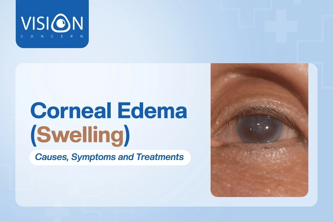
Eyes are sensitive; they are protected via a corneal membrane. What if it shows signs of redness or swelling? We talk about abnormalities today on how the epithelial layer, endothelium, and conditions like Fuchs' dystrophy or infections result in corneal edema.
Corneal edema can lead to a range of visual symptoms, like halos, and reduced glare, especially at night. Your cornea can appear cloudy and to be more precise about clinical features do consult your doctor. There are conditions that corneal swelling can progress into like
Initially, to fix any eye-related issue, diagnosis is key. Corneal edema is often diagnosed with,
| Procedure | Diagnosis |
|---|---|
| Slit-lamp Examination | Visual inspection to see signs of swelling, blisters, and haziness |
| Pachymetry | To identify swelling areas and measure corneal thickness |
| Endothelial Cell Count | Assess potential damage or dysfunction |
| Gonioscopy | Look out for signs of glaucoma that can worsen the case |
| Ultrasound | Assess the internal structure of the eyes and trace swelling |
| Laboratory tests | To check systematic conditions associated with corneal edema |
Your eyes can also swell like another part of your body when there is too much fluid inside. Corneal edema deals with abnormalities in corneal transparency. You know which to blame for this, corneal endothelium.
If anything is wrong there, it's obvious it won't be as transparent as in an ideal case. The endothelium is responsible for regulating the water content in and out of the eyes; there are many cases in which it cannot properly function.
It's a progressive but non-inflammatory condition where an endothelial layer of the cornea is impacted. There are four stages of this hereditary and progressive disorder, out of which, you see, no signs in stage 1.
Stage 2 denotes the situation when you have painless halos and vision loss around the periphery of the eyes. As stage 3 approaches, pain, and blisters become more noticeable, and even the edema protrudes out. Stage 4 can severely affect corneal opacity and may also result in permanent vision loss.
It usually is seen in individuals over 50, especially women, who face the loss of endothelial cells (which maintain transparency in the eyes). It is hereditary and a 50% chance that the patient inherits from their generation tree is seen. The remaining half of the cases are sporadic, and not seen in the family history.
When the cornea is inflamed, it fails to maintain hydration and corneal clarity. It in turn results in fluid deposition in the cornea and causes swelling. The active cause for this could be viral infections, like
Even when the body system shows an autoimmune response against the endothelial cells, such inflammation results. Use of tropical medication, procedural intervention in surgery, and exposure to irritants or toxins can cause inflammation in the corneal layer.
Such an inflammation causes the release of cytokines and mediators in irregular patterns, messing around with the ability to pump fluid out of the cornea. This can cause halos around lights, discomfort, and anterior chamber inflammation. This is why Vision Concern clinic pays active caution in clinical examination.
The swelling of the cornea is possible due to eye energy if not sought effective treatment within time. Your eyes could be showing signs of issues and injury; get it effectively diagnosed. The puncture wounds due to sharp objects can damage both the corneal and endothelial layers of the eyes.
Trauma to the eye can disrupt endothelial function. There are chemical burns, surgical trauma, thermal injuries, and blunt force trauma that can result in the severity of edema. Remember you are not alone; if your brother pokes you in the eye or some chemicals enter your remember, project, timely treatment can protect you from corneal swelling or prolonged chances of vision loss.
High intraocular pressure can often damage our optic nerve, which transmits our visual signal to the brain. When it grows more, one is diagnosed with glaucoma. This is not it; it causes vision loss and even triggers corneal swelling as the secondary effect.
The corneas are less resilient and more susceptible to damage because of the increased pressure. Optometrists measure the progression of glaucoma to see fluctuations in IOP for you to grant an appropriate treatment.
Accidents, surgery, and conditions like glaucoma damage the endothelial cell density and function. If you ask, endothelial damage is caused by more than one reason, like,
Endothelial damage is often associated with corneal dystrophies such as,
Prolonged use or irritation from contact lenses can contribute to corneal swelling. When the cornea retains more fluid, it is obvious to one to have vision impairment. The cornea mostly relies on oxygen and soft contact lenses have less oxygen permeability.
Wearing contact lenses for too long can irritate your eyes. It can be a real pain if use it for more than 6 hours a day. What’s hypoxic is they might be infected with bacteria and still be on our eyes for so long, increasing the chances of infection, scarring, and swelling of the cornea.
We know rheumatoid arthritis for joint pain, but this can cause pain in the eyes too. When our eye’s outer layer is inflamed, it triggers a condition called scleritis or episcleritis results.
If the cornea gets any thinner they are vulnerable to endothelial cell loss. Before your vision shows any concerns, having preventive strategies is the best-solved case. It helps catch problem early and reduce eye-related complications.
An inflammation like uveitis can lead to the release of harmful substances. This can impair the endothelial cell function and often cause edema.
Uveitis is something you dont know, there is this layer of tissue on the back of the eye called the uveal tract, that sometimes malfunctions. It's the same condition we are talking about, when it happens, it releases cytokines, that are not good for our corneal health. It even escalates to trauma for the eyes if not controlled.
Even bacteria and fungi can attack our eyes, and affect the endothelial cells. Endophthalmitis can be a medical emergency as it impairs the endothelium function and results accumulation of harmful substances on the cornea.
Corneal graft infection is no exception. There are risk factors like microbial contamination, ocular surface disease, and suture-related issues. Usually, pathogens like viruses worsen the case after a corneal transplant procedure, which is not any pleasant for our eyes,
There are different cases of corneal swelling, treatment varies from stage to stage. If the inflammation is caused due to glaucoma, it is suggested that one to,
Treatment options
| Condition | Treatment Options |
|---|---|
| Corneal dehydration | Hypertonic agents (5% NaCl anhydrous glycerine) |
| Pain relief for corneal edema | Hot forced air from a hair dryer (temporary relief) |
| Bullous keratopathy | Use of therapeutic soft contact lenses |
| Severe corneal edema | Penetrating keratoplasty (corneal transplant) |
| Mild corneal edema | Saline eye drops |
| Severe corneal edema with vision issues | Corneal transplant or DSEK surgery |
As the research made by suggests, the normal water content of the cornea is 78%, but the hydration is more than that, and the central thickness of the cornea increases. It causes the transparency to reduce, as the water pumps the action of the corneal endothelium.
In normal cases, the pressure (swelling one) is constant by the balance of the stromal matrix. 60 mm of Hg can act as a barrier action to prevent corneal swelling. There are reasons for abnormalities like,
It may not always be possible to prevent corneal swelling. But we can be cautious from our end in treating the case. In that case, regular eye exams and diagnosis of underlying conditions can help reduce the risk. We can treat it further with the doctor’s help. For a trusted clinic in Kathmandu, you can always refer to the Vision Concern clinic.
Yes, we provide emergency eye care for conditions like eye injuries, sudden vision loss, and infections. If you experience any urgent eye problems, please contact us immediately, and our team will assist you in getting the care you need.
Signs to watch for include blurry vision, floaters, sudden loss of vision, eye pain, redness, or sensitivity to light. If you experience any of these symptoms, it’s important to schedule an eye exam at Vision Concern Eye Clinic as soon as possible for early diagnosis and treatment.
If you’re experiencing blurred vision, headaches, or eye strain, it may be a sign that you need glasses or contact lenses. Our eye exams will help determine whether you need corrective lenses. We’ll also discuss your options based on your lifestyle and preferences, including glasses, contacts, or even refractive surgery like LASIK.